
A pressure sore is not limited to what is shown on the skin’s surface. The actual depth of tissue damage, the presence of a subcutaneous detachment, or bone contact systematically escape photographic observation. Here we discuss the progression of pressure sores from stage 1 to stage 4, emphasizing what the image reveals, and especially what it conceals.
Standardized Photography Protocol for Monitoring Pressure Sores
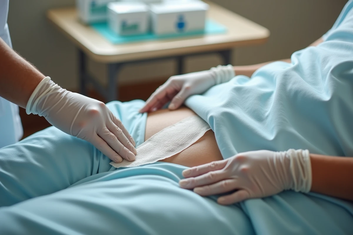
A photo of a pressure sore has clinical value only if it meets specific technical conditions. Too many shots taken with a smartphone in yellow artificial light, at varying angles, make any comparison impossible from one week to the next.
See also : Discover the secret of yellow wine chicken Robuchon style to elevate your meals
The criteria now considered minimal for usable remote monitoring are strict:
- Fixed distance between the camera and the wound, with a graduated scale placed at the same level as the lesion to allow for reliable measurement of dimensions
- Diffuse non-directional cold light, which avoids shadows that distort the assessment of depth and color of the wound bed
- Strictly perpendicular capture to the skin, against a neutral background, to limit geometric distortions
Without these conditions, comparing two photos taken a week apart is akin to comparing two different images of the same reality. The impression of improvement or worsening can be a mere lighting artifact.
See also : Discover how to log in to the student area of the Rouen ENT in no time!
We recommend pairing each photographic series with a structured collection: measured dimensions with a ruler, description of the wound bed (percentage of fibrinous, necrotic, granulating tissue), volume of exudate, odor. These textual data compensate for the inherent limitations of the image.
A visual guide presenting photos of pressure sores from stage 1 to 4 remains useful as a comparative reference for caregivers, provided it is never used as a standalone staging tool.
Stage 1 and Stage 2 Pressure Sores: What Redness Does Not Reveal
At stage 1, the lesion manifests as a persistent redness that does not blanch under finger pressure. The skin remains intact. On dark skin, this redness may take on a purplish or brownish hue, making detection significantly more difficult in photos.
The temptation is to consider this stage as benign. In reality, the redness indicates already established tissue ischemia. The absence of skin breakdown does not exclude suffering in the deeper layers. Perilesional palpation, which cannot be assessed in a photo, provides critical information: induration, local warmth, provoked pain.
At stage 2, tissue loss reaches the dermis. The wound may present as a serous blister or a superficial abrasion. Visually, distinguishing between stage 2 and moisture dermatitis (a lesion related to maceration) regularly poses problems, even for experienced caregivers. The precise location on the pressure area, the shape, and the clinical context guide the diagnosis more than the photo alone.
Stage 3 and Stage 4: The Invisible Depth in an Image
Stage 3 marks the transition to complete tissue loss reaching the hypodermis. Subcutaneous fat may be visible in the wound bed, but the muscle fascia remains intact. On a pressure sore, the sometimes abundant adipose tissue in this area can give the impression of a moderate cavity, while the subcutaneous detachment extends well beyond the visible edges.
It is precisely at stage 3 that the photo becomes the most misleading. The skin opening, sometimes narrow, does not reflect the actual extent of the underlying pocket. Only a probing examination allows for exploration of the depth and the existence of fistulous tracts.

At stage 4, the destruction reaches muscle, tendon, or bone. On the buttock, bone contact with the sacrum or ischium indicates a stage 4 injury and suggests a risk of osteitis. The photograph often shows a striking wound, with black necrotic tissue or exposed anatomical structures. In reality, even at this stage, the image frequently underestimates the severity: necrosis can cover and mask the actual depth of the ulceration.
The SFFPC reminded during the 2024 Healing Days that the photo is only a complementary tool for remote monitoring and should never serve as the sole basis for deciding on a significant intervention such as surgical debridement or intravenous antibiotic therapy.
Reliable Assessment of a Pressure Sore: Beyond the Snapshot
A structured clinical assessment systematically combines several dimensions that photography does not capture. Field feedback on wounds and healing converges on a minimal foundation:
- Perilesional palpation to detect induration, fluctuation, subcutaneous crepitus
- Button probe test to explore depth and check for bone contact
- Assessment of pain (spontaneous and provoked), odor, and volume of exudate
- Consideration of nutritional context and comorbidities (diabetes, arterial disease) that radically alter the healing prognosis
An undernourished patient with a stage 3 pressure sore may have a darker prognosis than a well-nourished patient with a localized stage 4 sore. Visual staging alone does not allow for this prioritization.
Photographic Monitoring in Daily Practice
Photo monitoring remains relevant for objectifying changes over time, provided the standardized protocol is followed and integrated into a complete care record. In facilities as well as at home, the series of timestamped images allows different caregivers to share a common visual basis.
The image documents, it does not diagnose. We regularly observe situations where a reassuring snapshot masks a deep worsening detectable only through physical examination. Conversely, a visually striking sore at the debridement stage may correspond to a favorable evolution.
The staging of a pressure sore remains a comprehensive clinical act. Any therapeutic decision based solely on a photograph exposes one to management errors whose consequences, at this anatomical level, can include difficult-to-reverse bone complications.